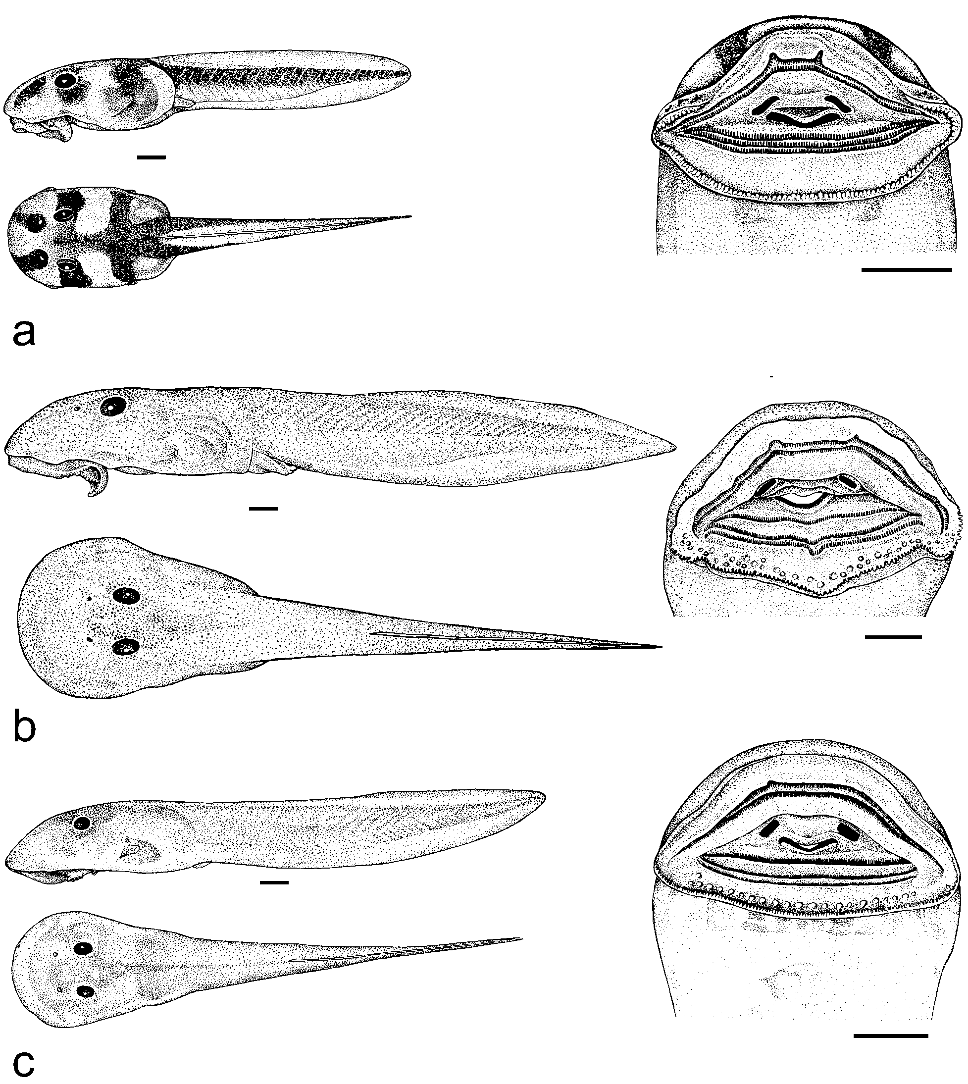
Epidermal melanocytes are small, approximately circular, with irregular fringes. The venter and oral disc are mostly unpigmented and translucent, except for the lateral parts at the transition to the flanks (Fig. 6 b). The gills and the heart shine through the ventral skin in red; developing front limbs are visible through the skin. The gut coil is arranged spirally and clearly visible in ventral view (Fig. 6 b). The iris is black with a clear golden ring around the pupil. Pigmentation is absent on the oral disc. In general the tail blood vessels are not conspicuous, but the vena caudalis ventralis is visible in ventral view. The ventral side of the muscular part of the tail is devoid of pigmentation. Tail fins are with light pigmentation with increasing melanocyte density toward the margins of the fin. The colour in preservation is similar to colouration in life, just lighter. The golden sheen of the described clusters of cells and the golden rim of the iris disappears upon preservation. External morphological features. Ansonia minuta is a small to mid-sized tadpole. The maximum total length in our sample was 23.7 mm at stage 39 (Tab. 2); the tail accounts for 61 – 64 % of total length. In dorsal view the body contour is pear shaped with a slight constriction behind the level of the eye (Fig. 4 b, 6 a). The body is widest anterior to the eyes. The body is dorsoventrally depressed. The snout is greatly expanded bearing a large sucker (oral disc) ventrally. In life and adhering to the substrate, the snout profile is convex (Fig. 4 b, 6 c). The eyes are located dorsally, at clear distance from the body contour in dorsal view. The external nares are much closer to the eyes than to the snout (Fig. 6 a). The nares have a smooth rim without projections. The interorbital distance is 1.25 ± 0.14 (mean ± SD) times the internarinal distance. The spiracle is sinistral and the spiracular orifice is attached to the body wall medially, free laterally. The spiracle is low on the flank in lateral view when the tadpole is attached to a rock (Fig. 6 c). The spiracle is directed posterodorsally. The oral disc is ventral and as wide as the snout in adhesion state (Fig. 6 b). An uniserial row of marginal papillation is present on the lower lip of the oral disc; the margin of the upper lip lacks papillae (Fig. 4 b). The oral disc margins possess inconspicuous lateral indentations at the end of the papillae row. Papillae are short, blunt and adjoining. Flat, rounded submarginal papillae are present on the lower lip: one complete posterior row (18 – 21 papillae) and an incomplete anterior one (3 – 6 papillae on each side; gap medially). The labial ridges bear uniserial keratodont rows. The Labial Tooth Row Formula (LTRF) is 2 / 3. The upper lip keratodont rows reach the level of the lower lip keratodont rows caudally (Fig. 6 b). The keratodonts are spoon-shaped with incisions along their edges (Fig. 6). Keratodonts are relatively more finely serrated on the posterior than on the anterior lower lip rows (Fig. 7). Keratodonts on the two upper lip rows are less clearly differentiated (Fig. 7 a – b). The upper beak comprises two short, widely spaced beak edges; the gap is 2.4 – 3.2 x the length of one beak edge. The lower beak is shallow V-shaped and serrated along its edge. The tail musculature is strong and almost as high as the trunk (in lateral view) at the trunk-tail junction. It reduces in height distally with a smooth but significant reduction in height at about mid-tail length. The dorsal tail fin starts only at ca. 40 % of the tail length. Dorsal and ventral tail fins are of approximately equal heights. The tail reaches its maximum height at mid-tail position. The edges of the fins edges are only slightly convex in lateral view, the shape is spatulate, tapering to a narrowly rounded tip (Fig. 6 c). The anal siphon is located medially. Variation. The described pigmentation features did not vary among specimens examined, however, individuals may vary in the densities of golden pigment cells, and we do not know if early larval stages have them. The anterior rise of the tail fins was found smoother in some specimens than in the individual in Fig. 6 (see Tab 2 for metric variation). Ecological notes. We collected Ansonia minuta tadpoles from rock faces in strong, foaming (presumably high energy) current. Tadpoles appeared active during the day and also by night. We observed them on a spectrum of rock inclinations, from flat to vertical, but always in strong current.