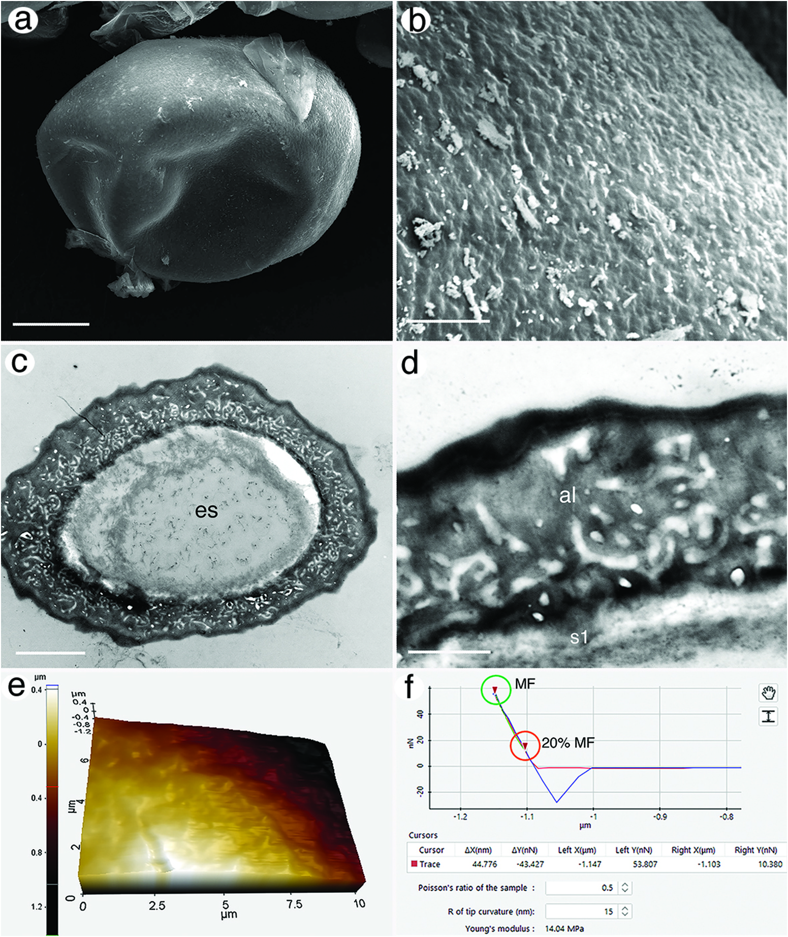
SEM Diapausing embryos were ovoid and measured ca 134 µm in diameter (̄ x = 134.0 ± 4.0 μm). The outer surface was composed of numerous smooth ridges and wrinkles, creating an overall rough surface (Figure 2 a, b). TEM The shell consisted of a thick apical layer and a thin basal layer. Average shell thickness was 2.30 ± 0.22 µm (SD). The outer layer, termed the alveolar layer by Munuswamy et al. (1996), had a wavy texture (Figure 2 c, d). Electron density was highest at the topmost and bottom portions of the alveolar layer, otherwise it had a similar electron density throughout its thickness and contained many ̍ empty ̾ pockets that were interconnected through electron-dense bridges. The basal layer [s 1 of Munuswamy et al. (1996)] had an amorphous appearance that consisted of an electron lucent zone and a more electron-opaque zone below it (Figure 2 d). Thin fibres were present throughout both zones. AFM Topography of the outer shell showed rugosity and similar attributes to those in SEM, but with fewer details (Figure 2 e). An example force – distance curve that displays how Young ̾ s modulus values were collected for a single spot on one egg is shown in Figure 2 f. The Young ̾ s modulus ranged from 12.24 to 19.85 MPa with an average of 15.81 ± 2.49 MPa (SD) (Table 1). Hardness values ranged from 1.14 × 10 − 2 to 5.31 × 10 − 2 GPa with an average hardness of 1.853 × 10 − 2 ± 1.23 × 10 − 2 GPa (Table 2).
Sumber: Integrative microscopy to explore physical and nanomechanical properties of eggshells of diapausing embryos in Rotifera: a proof-of-concept study